Dr. Chen viewed Anderson, James
Platform Features
Everything you need for ophthalmic imaging
One platform to integrate, store, view, and collaborate on all your imaging data. Built for the way modern eye care practices actually work.
Platform Overview
What is Unified?
See why practices choose Unified to orchestrate their ophthalmic imaging workflow.
Enable subtitles for the full experience
Capabilities
Everything your practice needs
From image capture to clinical collaboration, powered by AI.
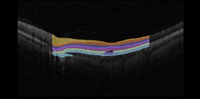
OCT
Fundus
Compare Images
OD Jul 16, 2025 NW400
OD Jan 10, 2025 maestro
Differences
Changes detected: 2 regions
Temporal
Inferior arcade
01/24/2026 • 4 images
01/10/2026 • 3 images
Retina Help
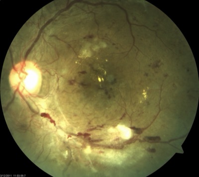
AI
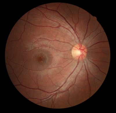
OD Fundus - Photography
DR Classification
No DR
92.00%
Mild NPDR
8.00%
Moderate NPDR
0.00%
Severe NPDR
0.00%
Virtual Scribe
AI
Source Image OD Fundus - Photography
Assessment
Mild non-proliferative diabetic retinopathywith scattered microaneurysms in the posteriorpole. No macular edema detected.
Plan
Follow-up in 3 months
Continue current management
Repeat OCT at next visit
AI Confidence: High
Reviewed & Approved
Custom Models
Beta
OD Fundus — Photography
Predictions
drusen
0%
no drusen
100%
Model Performance
Accuracy 94.2%
Training data 1,247 images
Last updated Jan 15, 2026
Available Models
Secure Message
HIPAA Re: Anderson, James — OCT Review
Type a secure message...
End-to-end encrypted
Cohort
Retina Case Review PHI Tracked
Content Full Patient
Members
RC
JP
SM
AK
LW
+3
Shared Cases
OD Fundus
OS Fundus
OCT B-scan
8 members
12 cases
Active
Patient Timeline
A
Anderson, James MRN 104829Jan 23, 2026 Annual Exam
4 images captured Dr. Chen
Oct 15, 2025 Follow-up
2 images captured Dr. Park
Jul 02, 2025 Screening
3 images captured Dr. Chen
Jan 10, 2025 Initial Visit
1 image captured Dr. Mills
Notifications 3 new
2m ago
14m ago
1h ago
3h ago
Yesterday
Dashboard
Patients Seen
284 +12.3%
Images Captured
1,247 +8.1%
Reports Generated
156 +5.7%
Avg Review Time
4.2 min -3.4%
Daily Activity
Clinical Report
AutoAnderson, James DOB 03/14/1978
MRN 40291Findings
Mild NPDR detected OD Monitor
Stable macular thickness OS Stable
IOP within normal range OU Stable
Export
Billing Codes
Auto-suggest Anderson, James — Jan 23, 2026
Suggested Codes
ICD-10 Codes
E11.319 Type 2 diabetes with mild NPDR
H35.30 Unspecified macular degeneration
Audit Log HIPAA
View
Dr. Patel edited Garcia, Maria diagnosis
Dr. Kim shared Thompson, Lisa with Dr. Park
Dr. Park exported OCT report
System auto-backup completed
Dr. Rivera viewed Williams, Sarah
847 events this month 12%
Last audit: 2 min ago Connected Devices 6 devices
5 Online
1 Offline
Topcon NW400 Fundus
Zeiss Cirrus 6000 OCT
Optomed Aurora Fundus
Heidelberg Spectralis OCT
Canon CR-2 Fundus
Haag-Streit BQ 900 Slit Lamp
Getting Started
4/6 complete
Welcome, Dr. Chen!
Let's set up your practice
Create your account
Verify email address
Set up your practice profile
Connect your first device
Invite team members
Import existing patients
Imaging
See Everything in One View
Multi-format viewing from OCT to fundus, side-by-side comparison, and a full patient gallery.
OCT
Fundus
Compare Images
OD Jul 16, 2025 NW400
OD Jan 10, 2025 maestro
Differences
Changes detected: 2 regions
Temporal
Inferior arcade
01/24/2026 • 4 images
01/10/2026 • 3 images
AI & Analysis
Intelligence Built In
AI-powered DR screening, automated clinical documentation, and custom model training on your own data.
Retina Help
AI
OD Fundus - Photography
DR Classification
No DR
92.00%
Mild NPDR
8.00%
Moderate NPDR
0.00%
Severe NPDR
0.00%
Virtual Scribe
AI
Source Image OD Fundus - Photography
Assessment
Mild non-proliferative diabetic retinopathywith scattered microaneurysms in the posteriorpole. No macular edema detected.
Plan
Follow-up in 3 months
Continue current management
Repeat OCT at next visit
AI Confidence: High
Reviewed & Approved
Custom Models
Beta
OD Fundus — Photography
Predictions
drusen
0%
no drusen
100%
Model Performance
Accuracy 94.2%
Training data 1,247 images
Last updated Jan 15, 2026
Available Models
Collaboration
Work Together Seamlessly
Instant image sharing, HIPAA-compliant messaging, and research cohort management.
Secure Message
HIPAA Re: Anderson, James — OCT Review
Type a secure message...
End-to-end encrypted
Cohort
Retina Case Review PHI Tracked
Content Full Patient
Members
RC
JP
SM
AK
LW
+3
Shared Cases
OD Fundus
OS Fundus
OCT B-scan
8 members
12 cases
Active
Patient Management
Every Patient Every Detail
Longitudinal timelines, smart notifications, and real-time dashboard analytics.
Patient Timeline
A
Anderson, James MRN 104829Jan 23, 2026 Annual Exam
4 images captured Dr. Chen
Oct 15, 2025 Follow-up
2 images captured Dr. Park
Jul 02, 2025 Screening
3 images captured Dr. Chen
Jan 10, 2025 Initial Visit
1 image captured Dr. Mills
Notifications 3 new
2m ago
14m ago
1h ago
3h ago
Yesterday
Dashboard
Patients Seen
284 +12.3%
Images Captured
1,247 +8.1%
Reports Generated
156 +5.7%
Avg Review Time
4.2 min -3.4%
Daily Activity
Reporting
Insights That Drive Action
Customizable reports, automated billing workflows, and complete audit trails.
Clinical Report
AutoAnderson, James DOB 03/14/1978
MRN 40291Findings
Mild NPDR detected OD Monitor
Stable macular thickness OS Stable
IOP within normal range OU Stable
Export
Billing Codes
Auto-suggest Anderson, James — Jan 23, 2026
Suggested Codes
ICD-10 Codes
E11.319 Type 2 diabetes with mild NPDR
H35.30 Unspecified macular degeneration
Audit Log HIPAA
Dr. Chen viewed Anderson, James
Dr. Patel edited Garcia, Maria diagnosis
Dr. Kim shared Thompson, Lisa with Dr. Park
Dr. Park exported OCT report
System auto-backup completed
Dr. Rivera viewed Williams, Sarah
847 events this month 12%
Last audit: 2 min agoIntegrations
Connects to Everything
Works with all major imaging devices, plug-and-play onboarding, and open APIs.
Connected Devices 6 devices
5 Online
1 Offline
Topcon NW400 Fundus
Zeiss Cirrus 6000 OCT
Optomed Aurora Fundus
Heidelberg Spectralis OCT
Canon CR-2 Fundus
Haag-Streit BQ 900 Slit Lamp
Getting Started
4/6 complete
Welcome, Dr. Chen!
Let's set up your practice
Create your account
Verify email address
Set up your practice profile
Connect your first device
Invite team members
Import existing patients
Cross-Platform
Works Everywhere
Access your imaging data from any device — a full-featured web app at your desk, and a native iOS experience on the go.
Web App
Full-featured platform accessible from any browser. Manage patients, view images, run AI analysis, and collaborate — all from your desktop.
Dashboard
Patients Seen
284 +12.3%
Images Captured
1,247 +8.1%
Reports Generated
156 +5.7%
Avg Review Time
4.2 min -3.4%
Daily Activity
Fundus
iOS App
A native mobile experience built for clinicians on the go. Capture, review, and share images with an interface designed for touch.
Good afternoon, Dr. Jane Demo UI demos
Recent Patients
SG
Sherry Gann
BS
Benjamin Swift
AL
Ava Lindstrom
Images by Modality
69
Photography
Ophthalmic Ph.
Custom
OCT
Slit Lamp
Clerical
Topography
Visual Field
Other
Integrations
Works with your existing tools
Seamless integration with all major imaging manufacturers, EHR systems, and AI analysis tools.
AI Partners
- RHRetinaHelpFundus DR screening
- ALAltrisOCT biomarker detection
All Major Devices
Works with virtually every ophthalmic imaging device including:
Zeiss Topcon Heidelberg Optovue Canon Nikon + more
EHR Systems
Seamless integration with major electronic health record systems:
Epic Cerner Athena NextGen + custom
Ready to see the whole
patient story?
Join 500+ eye care locations already using Unified to transform their imaging workflow. Get started with a personalized demo.